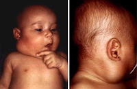
The most common diagnostic tools used to investigate the effect is lymphangioma and ultrasound, Doppler ultrasound, computed tomography (CT scan) and magnetic resonance imaging. Ultrasound shows hypoechogenic superficial multilocular cystic mass, but failed to show expansion retropharyngeal, axillary or mediastinum. With a CT scan, it may be difficult to distinguish the mass of soft tissue structures. More recently, MRI has been shown to be better in describing the boundaries of tumors and can more accurately describe the network.
Medical Health Information about Etiology, Clinical Manifestations, Diagnosis, Treatment and Therapy of Disease also Healthy Tips
Lymphangioma Signs and Symptoms
 |
| Lymphangioma |
Lymphangioma Signs and symptoms vary according to the anatomical location. Two-thirds of cervical lymphangioma showed no symptoms. They are present as swelling multilobular, palpable cystic on palpation. Massa may have a limit but most often infiltrate. Posterior triangle of the neck and submandibular region was the most common being the location presentations.
Lymphangioma
 |
| Lymphangioma |
Definition
Lymphangioma is a collection of abnormal benign lymphatic vessels that form a mass that consists of cystic spaces with the size of the variance, which have the potential for local extension, so it can infiltrate surrounding structures. Lymphangioma, cystic hygroma, and limfangiomatosis can affect almost any area of the body where there is lymphatic tissue, but mostly in the head, neck and axilla.
Cyst of Gartner
1. Definition
Gartner cyst with other names Gartner duct cyst or cysts Gartnerian, is vaginal cystic tumors are benign, coming from the rest of Gartner duct (duct epoophoron longitudinal) or the embryonic mesonephros and Wolffian duct system. These cysts arising from the terminal duct Wolffian that
develops as a result of the blockage of the duct secretions produced.
Gartner duct cysts and a thin-walled translucent consisting of epithelial stratified squamous or columnar epithelium or may be both. These tumors usually found in the vaginal wall and rarely occurs in the area of the labia minora clitoris or the hymen.
Steaocystoma Multiplex
Synonyms
Sebocystomatosis, Steatocystoma Multiplex Pringler
Definition
Steatocystoma multiplex is a typical disease, characterized by the presence of multiple cysts dermis, which contains sebum, and limited by the epithelium containing sebaceous follicles. Autosomal dominant inherited.
Epidemiology
It usually begins at puberty or young adulthood. Men more often affected than women.
Etiology
Formerly considered a keratin or sebaceous cysts inclusion. Now steatocystoma considered a hamartoma and variant of the cyst or dermoid cyst vellus hair.
Clinical manifestations
Lesions can arise at birth or shortly afterwards. Cystic nodules appear clinically asymptomatic with soft consistency up hard, attached to the overlying skin, yellowish with surface smooth and when the lesion is punctured will discharge oily yellow like cheese. Its size varies, from a few mm to 3 cm, but rarely more than 1.5 cm. In general, the lesion is located in the sternum area, axillary arm, and scrotal area.
Histopathology
In microscopic, cyst appear as layer of squamous epithelium without granular layer. Cyst usually folded. Characteristics that appear as non cell layer thick form, eosinophilic, homogeneous, epithelial lining lumen side. Cyst wall often consists of adnexal structures primarily glandular sebaceous or abortive hair follicles. Cyst space can contain lanugo hair.
Differential diagnosis
Dermoid cysts, epidermoid cyst multiple, Neurofirbomatosis, sebaceous adenoma, Lipoma.
Treatment
The best treatment is by excision. But because there are many, such methods sometimes can not be implemented. As an alternative incision and the contents of the cyst expression, but it is often cause recurrence after a few months
MILIUM
Definition: Milium a keratin cyst subepidermal small, especially occur on the face, especially periorbital. Originating from the epidermis or adnexal, may occur secaa primary or secondary.
Epidemiology: Often found in the parent, but may occur in infants newborn. More common in women than men.
Etiology: The cause of primary milia ridak known, is likely to come from pilosebaseus follicles. While the secondary milia common of retention cysts after various dermatoses, ascribed to the hair follicles, glands sweat, sebaceous glands or the epidermis.
Histopathology: microscopic description similar to the epidermal cyst, only different in size. On a serial pieces, with primary milium Velus appear to be associated with a hair follicle, while milium
Secondary appear to be associated with formation of epithelial stem.
Differential diagnosis: Pustular Acne, Molluskum contagiosum, Hyperplasia sebassa.
Treatment: Incision and contents milium expression. Prognosis: Milia are purely benign lesions and just cause cosmetic problems.
Trichilemmal Cyst
Synonyms: Kista pilaris, sebaceous cysts.
Definition: trichilemmal cyst is a cyst containing keratin, composed by an epithelium that resembles the outer root sheath of hair, can be derived autosomal dominant
Epidemiology: Usually appears in middle age. Women are more often affected than men.
Etiology: Wall cysts originating from outside the hair root sheath that surrounds the bottom of the hair follicle.
Manifesasi Clinic: basanya occurs on the scalp. Clinically difficult to distinguish from epidermal cysts, but these cysts dienukleasi easier and more keratinosa contents and not so fatty and less smelly than the content of epidermal cysts.
Histopathology: Looks restricted cyst wall several layers of cuboidal shaped epidermal cells are arranged palisade without granular layer and intercellular bridges. Epithelial cells bordering the cyst contents swell and contains pale cytoplasm. Cyst contents in the form of a homogeneous eosinophilic material.
Diagnosis: epidermal cysts, Cylindroma, Lipoma.
Treatment: Treatment for cysts trichilemmal is the same as epidermal cysts.
Prognosis: Good, rarely undergo malignant transformation.
Pathophysiology, Histopathology and Treatment of Epidermal cysts
Pathophysiology
Epidermal cysts occur as a result of the proliferation of epidermal cells in the dermis circumscribed space. In the analysis of epidermal cysts, lipid structure and the same pattern as in the cells of the epidermis. Epidermal cyst express cytokeratin 1 and 10. The source of the epidermis is almost always from the infundibulum of hair follicles.
Inflammatory mediated by section epdiermal keratinized cysts. In the study, the extract keratin is chemoactive for PMN.
The studies mentioned HPV (Human Papilloma Virus) and UV exposure plays a role in the formation of epidermal cyst.
How to change the epidermal cysts become cancerous is not known for certainly (though rarely epidermal cysts develop into malignant tumors). At epidermal cyst carcinoma, immunohistochemistry for HPV negative results, which can be summed HPV does not affect changes into squamous cell carcinoma. Chronic irritation and repetitive trauma to the limits of the cyst epithelium epidermis role in malignant transformation, but how to do is still unknown.
Histopathology
On histopathologic examination, epidermal cysts lined with stratified squamous epithelium containing granular layer. Laminated keratin found in cysts. Inflammatory responses can be found in the cysts rupture. Mature Cyst can be calcified.
TREATMENT
Generally, epidermal cysts do not require any treatment. When the excised can cause interference, or dissection entire cyst wall with incision. When part of the wall behind, the cyst may recur. Destruction of the cyst with curettage, liquid chemical, or electrodesiccation give unsatisfactory results.
When inflammation occurs, it can be given triamcinolone intralesional injection (amcort, aristocort) which can suppress PMN migration and make a narrow slit capillary blood vessels. Oral antibiotics are also given if necessary.
COMPLICATIONS
Very rare complications, including infection, scarring on removal, and recurrence. Malignancy in epidermal cysts are very rare.
Epidermoid Cysts
 |
| Epidermoid Cyst |
DEFINITIONS
Epidermoid cysts or also called a sebaceous cyst is a collection of materials such as keratin, usually white, slippery, easily moved, and cheesy on the inside wall of the cyst. This type of cyst is the most common. Clinically, epidermal cysts appear as rounded nodules, hard-colored flesh. Epidermal cysts generally have a small hole associated with the skin but are not always apparent.
Acrochordon Skin Tags
 |
| Acrochordon Skin Tags |
Treatment and Therapy of Seborrheic Keratosis
A. Drug Therapy
keratolytic agent
Can cause epithelial gore becomes fluffy, soft, maceration then desquamation.
1. Ammonium lactat lotion
Lactic acid and alpha hydroxy acids that have a keratolytic and keratin cells facilitating release . Perfomed 15% and 5% strenght; 12% strenght can cause irritation to the face for making keratin cells do not undergo adhesion.
2. trichloroacetic acid
Burn the skin, keratin and other tissues. May cause local irritation. Seborrheic keratosis treatment with 100% trichloroacetic acid can remove the lesion, but its use must be performed by professionals skilled hands .
Therapy can be used topical tazarotene 0.1% cream smear 2 times a day in 16 weeks showed improvement sheboroic keratosis in 7 of 15 patients.
Diagnosis of Seborrheic Keratosis
How To Make Diagnosis Seborrheic Keratosis
Diagnosis obtained through history and physical examination and investigation in the form of histology. Not necessary laboratory tests and radiological examination.
1. History
• Usually asymptomatic, patients complain of a lump of black, feels uncomfortable.
• lesions can sometimes itchy, wanted carded or flops.
• Patients sometimes feels a lump growing slowly.
• Lesions can not heal themselves suddenly.
• The majority of cases there is a family history of inherited.
• Lesions can occur throughout the body except the palms and soles, and mucous membranes.
Pathophysiology and Clinicopathologic Variants of Seborrheic Keratosis
Pathophysiology
Epidermal Growth Factor (EGF) or its receptor, has been shown to be involved in the formation of seborrheic keratosis. No significant differences of immunoreactive growth hormone receptor expression in epidermal keratinocytes in normal and seborrheic keratosis.
Seborrheic Keratosis
 |
| Seborrheic keratosis |
Seborrheic keratosis is a benign skin tumors that arise in most older people, about 20% of the population and are usually absent or rare in people with middle age. Seborrheic keratosis has many clinical manifestations can be seen, and seborrheic keratosis is formed of proliferation of epidermal cells of the skin. Seborrheic keratoses can appear in many forms lesions, lesions can be one or many types of lesions or multiple.
Trichoepithelioma
 |
| Trichoepithelioma |
Trichoepithelioma is a benign follicular tumors pilosebaseus apparatus may take the form of solitary or multiple. This tumor was first proposed by Brooke in 1892 under the name Epithelioma cysticum adenoids. Multiple Trichoepithelioma usually autosomal dominant inherited. While usually non-hereditary solitary form. These lesions begin to appear in childhood and will settle and multiply as adults. Generally found more than in women than in men.
Syringoma
 |
| Syringoma |
1. Syringoma periorbital (Periorbital Syrigoma)
2. Syringoma eruptive (Eruptive syringoma, Eruptive hidradenoma, Disseminated syringoma)
3. Another variant: the linear form of unilateral or nevoid distribution, linear finite, limited to the scalp, limited to the vulva, limited to the distal extremity, lichen planus-like, the type of milia (milia like).
Xanthelasma
 |
| Xanthelasma |
Subscribe to:
Posts (Atom)