Clinical Manifestations of Guillain-Barré Syndrome

1. Weakness
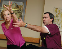
Classical clinical picture is ascending weakness and naturally symmetrical. Lower limbs are usually affected first before the upper limbs. Proximal musculature may be involved earlier than the more distal. Body, bulbar, and respiratory muscles can be affected as well. Respiratory muscle weakness with shortness of breath may be found, developed acute and lasts for few days to weeks. Severity can range from mild weakness to tetraplegia with ventilatory failure.

Pathophysiology of Guillain-Barré Syndrome

       The mechanism of how the infection, vaccination, trauma, or other factors that precipitate the occurrence of acute demyelination in GBS is still not known with certainty. Many experts conclude that the nerve damage that occurs in this syndrome is through immunologic mechanisms. Evidence that the immunopathogenesis which is the mechanism that causes peripheral nerve injury in this syndrome are:

Etiology of Guillain–Barré Syndrome

        What are causes of Guillain–Barré Syndrome? Etiology is still not yet known with certainty and the cause is still a matter of debate. Some conditions / diseases that precede and possibly something to do with the Guillain–Barré Syndrome, among others:

Guillain-Barre Syndrome

        Guillain-Barre Syndrome (GBS) is an autoimmune neurological disease in rare cases, in which the immune system produces antibodies against its own nerve, resulting in damage of the nerve. Guillain-Barre syndrome is also called acute inflammatory demyelinating polyneuropathy that attacks the nerve roots both ventral and dorsal acute symptoms and lead to paralysis of the lower limbs begin and extends up the body and facial muscles.

Medical Treatment and Therapy of Myasthenia Gravis

Medical Treatment and Therapy of Myasthenia Gravis
MG Treatment
         Although there is no research on treatment strategies for sure, but myasthenia gravis is a neurological disorder most treatable. Anticholinesterase (acetylcholinesterase inhibitor) and immunomodulating therapy is the main treatment of myasthenia gravis. Anticholinesterase commonly used in the mild myasthenia gravis. Whereas in patients with generalized myasthenia gravis, an immunomodulatory therapy needs to be done regularly.

Diagnosis of Myasthenia Gravis

Diagnosis of Myasthenia Gravis

       A careful physical examination should be performed to establish the diagnosis of a myasthenia gravis. Muscle weakness can occur in many different degrees, usually descend the proximal portion of the body and symmetrical on both left and right limbs. Deep tendon reflexes are usually still in the normal range. Myasthenia gravis is usually always accompanied by a weakness in the facial muscles. Bilateral weakness of facial muscles will cause a mask-like face in the presence of ptosis, and horizontal smile.

Clinical Manifestations of Myasthenia Gravis

Clinical Manifestations of Myasthenia Gravis
Ptosis in Myasthenia Gravis
      Myasthenia gravis is characterized by fluctuating weakness in skeletal muscle and this weakness will increase when it is active. Patients will feel very weak muscles in the day and this weakness will be reduced if the patient rested. Usuallyinitial complaint is a specific muscle weakness, not ordinary  muscle weakness  and the condition deteriorated typically fluctuate for several hours. Not apparent in the morning and usually worsens as the day progresses. It also varies over several weeks or months, with exacerbations and remissions.

Pathophysiology of Myasthenia Gravis

    When an action potential moves to motorneuron and reaches the motor end plate, the molecules of acetylcholine (ACh) is released from presynaptic vesicles, through the neuromuscular junction and then will interact with the ACh receptor (AchRs) in the membrane postsynaptic. AchRs canals open, allowing Na + and other cations into muscle fibers and cause depolarization. Persistent depolarization will converge into one, and if the accumulated depolarization is large enough, a meal will trigger an action potential, which moves along the muscle fibers to produce a contraction.

Myasthenia Gravis

Myasthenia Gravis
       What is Myasthenia Gravis? Myasthenia gravis is an autoimmune disorder caused by the presence of autoantibodies specific to human acetylcholine receptor (ACHR), which is concentrated in the synapses of the neuromuscular junction post. Myasthenia Gravis (MG) is a chronic, autoimmune causes muscle weakness and muscle fatigue slowly but progressively. If the patient is at rest, it is not long before muscle power will be restored. The disease is caused by the disruption of synaptic transmission at the neuromuscular junction.

Medical Treatment and Therapy of Parkinson's Disease

Medical Treatment and Therapy of Parkinson's Disease
       How to treat patient with Parkinson's Disease? To know the answer, lets read the explain. Parkinson's disease is a degenerative disease, which develops progressively, and the cause is unknown, therefore its management strategies are
1) symptomatic therapy, to maintain the independence of the patient,
2) neuroprotection and
3) neurorestoration,
both to inhibit the progression of Parkinson's disease. These strategies are aimed at maintaining the life quality of patient.

Diagnosis of Parkinson's Disease

Diagnosis of Parkinson's Disease
       How to diagnose Parkinson's Disease? Doctors must to know it. There are ways how to diagnose this disease. The diagnosis of Parkinson's disease is confirmed by the following criterias:

1. Clinically
• Achieved 2 of 3 cardinal signs of motor disorders: tremor, rigidity, bradykinesia or
• 3 of 4 motor signs: tremor, rigidity, postural instability and bradykinesia.

Pathophysiology of Parkinson's Disease

Pathophysiology of Parkinson's Disease
       In general, it can be said that Parkinson's disease occurs as a result of decreased levels of dopamine neuron death in the substantia nigra pars compact by 40 - 50% were accompanied by eosinophilic cytoplasmic inclusions (Lewy bodies). Lewy bodies are eosinophilic cytoplasmic inclusions with concentric peripheral halo and dense cores .

Clinical Manifestations of Parkinson's Disease

Parkinson's Disease clinical Manifestations
1. Motoric symptoms
a. Tremor
       Symptoms of Parkinson's disease often escapes ordinary sight, and seen as a normal thing in the elderly. One characteristic of Parkinson's disease is a hand tremor (shaking) when at rest. However, if the person is asked to do something, the vibe was not seen again. That's called resting tremor, which also lost during sleep.

Parkinson Disease

Parkinson Disease Mohammad Ali
Mohammad Ali
      If you look at the photo on the right, you can definitely tell a familiar face this. Yes, he is a former professional heavyweight boxer from the United States, Cassius Clay, better known by the name of the famous Mohammad Ali. In addition to the controversy, which had refused to join the military service, he also has Parkinson's disease. What is Parkinson's Disease?

Complications and Prognosis of Bell's Palsy

       If the disease Bell's Palsy is not treated immediately, it will gradually raises some complications. The following are the complications caused by Bell's Palsy.

Complications
a. Crocodile tear phenomenon
      That tears when the patient eats. These occur several months after the paresis and the consequences of incorrect regeneration of autonomic fibers are supposed to salivary glands, but to get to, lacrimal gland. Locations lesions around the geniculate ganglion.

Medical Treatment and Therapy of Bell's Palsy

Medical Treatment and Therapy of Bell's Palsy
       How to treat Bell's Palsy and Bell's Palsy patient care? A very good question and important to know. Here are the steps in the treatment of Bell's palsy.

A. Take Rest especially in the acute phase
The first therapy that is just enough rest during the attack or acute period. This is to rest the nerve and muscle paralysis.

Bell's Palsy Diagnosis

Bell's Palsy Diagnosis
      The diagnosis of Bell's palsy can be established by anamnesis and physical examination, in which case the neurological examination. To establish a diagnosis of Bell's palsy should be established first, the type of peripheral facial paresis, then get rid of all the possible causes facial paresis. Peripheral facial paresis is different, from the central type.

Clinical Manifestations of Bell's Palsy

Clinical Manifestations of Bell's Palsy
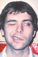
      Usually occurs suddenly, the patient is aware of the paralysis on one side of his face when they wake up in the morning, looked in the mirror or when brush teeth / rinse or told by another person / family that one corner of his face, lower. Bell's palsy is almost always unilateral. Clinical manifestations may include loss of all voluntary movement in paralysis.

Etiology and Pathophysiology of Bell's Palsy

Etiology and Pathophysiology of Bell's Palsy      Bell's palsy is idiopathic disease, and is a disease of the peripheral nerves, which are acute, and attacking the facial nerve (N.VII) that innervate all the muscles of the face, causing weakness or paralysis of one side of the face. This leads to the asymmetry of facial paralysis as well as disrupt normal function.        

Bell's Palsy

Bell's Palsy Medical therapy
         Facial paralysis is a form of disability which gives a strong impact on a person. Paralysis of the facial nerve can be caused by congenital birth (congenital), neoplasm, trauma, infections, exposure to toxic or iatrogenic causes. The most frequent cause of unilateral facial paralysis is Bell's palsy. Bell's palsy was found by a British physician named Charles Bell.
       

Post-Acute Phase Therapy of Stroke

Post-Acute Phase Therapy of Stroke
        After the acute phase passes, goals of treatment, emphasis on patient rehabilitation measures, and prevention of recurrence of stroke.

Preventive therapy
The aim is to prevent the recurrence of stroke or the onset of a new attack, by among others treat and prevent stroke risk factors:

Treatment and Therapy of Stroke Acute Phase

Treatment and Therapy of Stroke Acute Phase
       Therapy is divided into two phases, namely the acute and post-acute phase. Acute phase (0-14 days after onset of disease). Treatment goal is to rescue neurons that had not to die, and that other pathological processes that accompany not disturb / threatening brain function. Actions and drugs given should ensure perfusion of blood to the brain remained adequate, it does not reduce.So that needs to be maintained optimal function of respiration, heart, blood pressure, blood is maintained at optimal levels, control blood sugar levels (high blood sugar levels are not lowered drastically), when severe fluid balance, electrolyte, and acid-base should continue to be monitored.

How to Diagnose Stroke

How to Diganose Stroke
       Stroke is a medical emergency condition. Any person suspected of having a stroke should be immediately taken to a medical facility for evaluation and treatment. First, the doctor will ask the patient's medical history if there are danger signs ahead of time and do a physical exam.

Complications of Stroke

Complications in stroke Medical treatment therapy
       Complications in stroke often occur and cause clinical symptoms of a stroke, become increasingly worse. Signs of complications should be identified early so that it can be prevented from getting worse and to determine appropriate therapy. Complications in stroke are:

Clinical Symptoms of Subarachnoid Hemorrhage Stroke

Clinical Symptoms of Subarachnoid Hemorrhage Stroke
      Subarachnoid hemorrhage is commonly caused by rupture of an intracranial aneurysm. Before rupture, aneurysm usually does not cause symptoms until the press on nerves or blood leaking in small amounts, usually before a large outbreak (which causes headaches). Then generate an alarm, such as the following below:

Clinical Symptoms of Intracerebral Hemorrhage Stroke

Clinical Symptoms of Intracerebral Hemorrhage Stroke
    Symptoms caused by the onset of intracerebral hemorrhage that almost always arise during the move and sometimes occurs when the patient is sleeping (only 3%). The most common symptoms are headache and vomiting. Although not specific and dependent location of the lesion, it distinguishes ischemic stroke. Headache at onset is an important clinical symptom in patients with lobar hemorrhage, caused due to local distension, distortion, or stretching superficial intracranial structures that are sensitive to pain.

Hemorrhagic Stroke

Hemorrhagic Stroke Medical treatment therapy
Hemorrhagic Stroke
      Hemorrhagic stroke is a stroke caused by non-traumatic intracranial hemorrhage. In a hemorrhagic stroke, blood vessel rupture thereby inhibiting normal blood flow and blood seeping into an area in the brain and damage. Nearly 70% of cases of hemorrhagic stroke in patients with hypertension. Hemorrhagic strokes include bleeding in the brain (intracerebral hemorrhage) and bleeding between the inner and outer layers of tissue that protects the brain (subarachnoid hemorrhage). Other disorders which include bleeding within the skull, including epidural and subdural hematoma, which is usually caused by a head injury.

Clinical Symptoms of Ischemic Stroke

Clinical Symptoms of Ischemic Stroke medical treatment and therapy
        Clinical symptoms of ischemic stroke can occur at different locations depending on the neuroanatomical and vascularization were attacked, among others:

1. Anterior cerebral artery
         Anterior cerebral artery is an artery that provides blood supply to an area of the cerebral cortex parasagital, which includes motor and sensory cortex area for the contralateral limb, is also the center of inhibitoris bladder (micturition center). 

Ischemic Stroke

Ischemic Stroke Medical treatment and therapy
Ischemic Stroke
       Stroke Ischemic stroke is also called obstruction, or myocardial stroke, because of the events leading to decreased blood flow or even stop at a particular area of the brain, such as the occurrence of embolism or thrombosis. The decrease in blood flow causes the neurons stop functioning. Blood flow is less than 18 ml/100 mg / min will result in neurons that are irreversible ischemia. Most of the patients or 83% had this type of stroke.

Types of Stroke


Types of Stroke Medical Treatment Therapy
   Broadly based on the pathological abnormalities occur, a stroke can be classified as ischemic stroke and hemorrhagic stroke (bleeding). In ischemic stroke, blood flow to the brain stops because of atherosclerosis or a blood clot that has clogged the arteries. In hemorrhagic stroke, blood vessel rupture thereby inhibiting normal blood flow and blood seeping into an area in the brain and damage.

Causes and Risk Factors of Stroke

Causes and Risks Factors of Stroke medical treatment and therapy
         What are causes and risk factors of Stroke? nice question and we must to know the answer. Causes of stroke include atherosclerosis (thrombosis), embolism, hypertensive intracerebral hemorrhage and rupture causing saccular aneurysm. Stroke is usually accompanied by one or several other diseases such as hypertension, heart disease, increased blood fats, diabetes mellitus or peripheral vascular disease.

Definition of Stroke

A Stroke Patient get rehabilitation Medical Treatment and therapy
A strokepatient get rehabilitation
Definition of Stroke
      Stroke is the clinical term for non-traumatic disorders of blood circulation that occurs acutely in a focal area of the brain, which results in a state of ischemia and neurologic dysfunction, focal or global, which lasted more than 24 hours, or directly cause the death. In a matter of seconds and minutes, brain cells do not receive adequate blood flow more will die through a variety of pathological processes.Typically, stroke manifest as neurological deficits appearance suddenly, such as weakness or paralysis of movement, sensory deficits, or speech impairments may also.
Related Posts Plugin for WordPress, Blogger...