 |
| Pathophysiology Huntington's Disease |
Sensory information will be integrated at all levels of the nervous system and causes a motor respons , which began in the spinal cord, with the reflexes of the muscles are relatively simple, extends to the brain stem with a more complex response, and finally, extends to the cerebrum, where muscle efficiency the most complex controlled .
Stimulus motor can be divided into two, namely the pyramidal motor and extrapyramidal motor. Pyramidal motor centrum found in the frontal lobe of the cerebral cortex, which is the fourth area called precentralis gyrus or primary motor area, area 6 & 8 , known as premotoris area and area 44 & 45 or Broca's area that serves as a motor speech centrum .
Extrapyramidal motor centrum consists of cortex cerebri in areas 5 and 7 parietal lobe , area 22 in the temporal lobe, and area 19 on lobe occipital, ganglion basale, subthalamic, nucleus ventral and centromedianus / intralaminaria, thalamic reticular, cerebellum, nucleus ruber, substantia nigra, reticular formation, nucleus.
The role of the pyramidal motor is started or trigger the desired movement, producing " patron movement " is desirable, but the resulting motion is not smooth, yet skilled, and agile . While the role of extrapiramidalis motor is smooth motion that has been connected by the pyramidal motor so that the result of the interconnection of the two will result in regular movement, agile / skilled, and well coordinated .
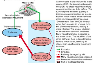
At Huntington neuron degeneration occurs in the cerebral cortex and basal ganglia to target neurons in the striatum, particularly those located in the caudate nucleus, putamen and globus palidus. In addition to produce neurotransmitters, the basal ganglia to the cerebral cortex plays an important role in the coordination of motor impulses which are primarily coordinated inhibition to produce the desired movement, controlling the relative intensity of a separate motion, direction of motion, and parallel sequencing of multiple movements. Therefore if there are abnormalities in the basal ganglia both structural and functional will eliminate the inhibitory effects that can be found on clinical abnormal movements that are repetitive or rhythmic. In line with progressive disease and chromosomal defects, will lead to the emergence of events that can lead to loss of neurons deficiency decarboxylation of glutamic acid and choline in the basal ganglia, causing biochemical disorder such as decreased production of GABA, the incidence of neuronal loss can also cause atrophy of the cortex which of course can lead to a lack of inhibition mechanism on fine motor pathway that controls coordination and smooth and agile movements become impaired can also manifest in the form of interference thought , perception and memory ( psychiatric disorders ).
Bilateral atrophy of the caudate nucleus caput region and putamen is a characteristic abnormality of Huntington 's disease, and generally also found gyrus atrophy in the frontal and temporal lobe regions. Atrophy of the caudate nuklelus result in changes in the appearance of the frontal horns formed on head CT scan images because of the lateral ventricles and sinisitra dextra, because the head of the caudate nucleus will give an idea prominent in the ventricle. In addition it will appear enlarged brain ventricles that go hand in hand with the disease progression .
Microscopically, degeneration happens is divided into 3 stages, early, moderately advanced, and far advanced. In the early stages, although it has been diagnosed by genetic testing, there is no striatal lesions, so that from this it can be concluded if the manifestations that arise due to biochemical abnormalities or infrastructural changes. This discovery is supported by the PET scan in patients with Huntington's disease in which glucose metabolism was found characteristic decrease in caudate nucleus which precedes the loss of tissue at an advanced stage. Striatal degeneration that occurred starting in the medial caudate nucleus and lateral spread to the are. Neuronal cells that exist in different sized brain and degeneration that occurs generally attack the neurons are small. Starting from the loss of dendrites of neurons are small, large neurons are generally not affected. Cells that undergo degeneration eventually be replaced by fibrous astrocytes. Anterior regions of the caudate and putamen are generally affected more extensively than the posterior region. Some researchers found a variety of changes in the globus pallidus, subthalamicus nuclei, Rubro nucleus, cerebellum, and substantia nigra pars reticucalata from. In areas of cerebral cortex, obtained neuronal loss was replaced by glial tissue .
 |
| Indirect pathway Huntington's Disease |
 |
| Direct pathway Huntington's Disease |
Mechanisms of the pathogenesis of Huntington's disease is an obvious but still hard to understand . Expansion of the region poliglutamine of Huntingtin ( Huntington gene product protein ) resulted in aggregation of the protein in the nuclei of brain neurons. Moreover, the protein has a tendency to aggregate in striatal neurons and cerebral cortex. The results of Wetz conclude if this protein is toxic to neurons directly or in the form that is not aggregated. But the problem lies there on the dominance of the Huntingtin protein aggregation, especially in regions of the cortex, whereas there is a loss of neurons in the striatal region. A theory is that if Huntington will cause certain neurons more sensitive to glutamate - mediated excitotoxicity. In addition, it is now proposed 2 mechanisms based on the interruption of transcription protein because the protein huntington protein binding to transcription, or mitochondrial dysfunction occur directly or through a similar transcriptional mechanism. Because poliglutamine expansion observed in various neurodegenerative disorders .
Source:
1. pict: http://dundeemedstudentnotes.wordpress.com/2012/10/07/pathology-and-neurobiology-of-huntingtons-disease/
2. pict: http://php.med.unsw.edu.au
3.http://www.medscape.org/viewarticle/759292
Related Articles
1. History and Etiology of Huntington's Disease
2. Clinical Manifestations of Huntington's Disease
3. Diagnosis and Differential Diagnosis of Huntington's Disease
4. Medical Treatment and Therapy of Huntington's Disease
Related Articles
1. History and Etiology of Huntington's Disease
2. Clinical Manifestations of Huntington's Disease
3. Diagnosis and Differential Diagnosis of Huntington's Disease
4. Medical Treatment and Therapy of Huntington's Disease
